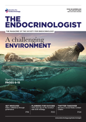
Exposure to endocrine-disrupting chemicals (EDCs) has been of growing concern to human and animal health over recent years.1 EDCs comprise a group of chemicals that can alter the release or action of endogenous hormones, and thus have the potential to alter normal physiological functions if they enter the body. Many EDCs are found in everyday items, such as packaging and food containers, personal care products, children’s toys, medical devices and pesticides, which end up in the environment (e.g. in surface or urban water and sewage) and are detectable in body tissues.
Human and animal exposure is ubiquitous. There is now a wealth of evidence to show that exposure to EDCs, particularly during gestational and pre-pubertal life, is implicated in testicular dysgenesis syndrome (TDS). This encompasses several male reproductive abnormalities of increasing prevalence, such as cryptorchidism, hypospadias, impaired semen quality and testicular germ cell cancer.2 Studying the effects of EDCs in humans is complex and has largely been limited to epidemiological studies. However, animal research has given a greater insight into our understanding of the role of environmental EDCs in declining male reproductive health.
ANIMAL STUDIES
Early evidence suggesting that exposure to EDCs may significantly affect the morphology of the male reproductive system in various species came from accidental exposures in wildlife, e.g. work by Edwards et al.3 In addition, several laboratory studies in rodents (some described below) have provided further substantial evidence that exposure to environmental EDCs can be detrimental to male reproductive health.
For example, rats exposed to benzo-a-pyrene (a chemical produced from incomplete combustion) exhibit reduced testis weight, lowered plasma and intratesticular testosterone concentrations, increased germ cell apoptosis, and decreased spermatozoa production.4,5 Similarly, prenatal exposure to bisphenol A, a chemical found in plastics, resulted in decreased sperm counts and motility in adult mice6 and disrupted meiotic progression during spermatogenesis in rats.7
‘Some studies have utilised a ‘real-life’ model of EDC exposure, using sheep exposed to pasture treated with biosolids.’
Other chemicals, such as the pesticide methoxychlor, have been shown to cause inhibition of testicular steroidogenesis in adult rats,8 which showed a reduction in proteins important for testosterone biosynthesis and production (steroidogenic acute regulatory protein, and 3β- and 17β-hydroxysteroid dehydrogenase). However, these effects were reversed after 72 hours.8 Another study in mice looked at gestational exposure to both di(2-ethylhexyl)phthalate (DEHP; a plasticiser) and polychlorinated biphenyls (formerly used in industrial manufacturing), which caused decreases in testicular weight, seminiferous tubule diameter, spermiogenesis, Leydig cell quantity, intratesticular testosterone, epididymal sperm count and sperm viability.9 Furthermore, in utero and lactational exposure to DEHP alone increased epididymal weight.10
Exposure to butylparaben, a chemical used in cosmetics, caused progressive spermatogenic cell detachment in rats that were orally exposed, and induced apoptotic cell death in the testes,11 as well as reductions in epididymal weight, sperm count and testosterone concentrations.12
While there is a lot of evidence linking effects of environmental exposure to EDCs and testicular dysgenesis in F1 generations, more recent evidence in mice has shown testicular changes in up to the third generation after maternal exposure. It is postulated that intergenerational effects of EDCs can occur via epigenetic DNA modifications.13 Though more research is required, this evidence could provide a deeper understanding of the longer term effects of EDC exposure in both human and animal populations.
REAL-LIFE MIXTURE EXPOSURES
Much of the research on EDCs and male reproductive function has utilised rodent models, due to their quick generation time, and has exposed them to high doses of single chemicals or limited mixtures of chemicals. However, such models do not replicate human chemical interactions, which involve chronic exposure to low level chemical mixtures in an outbred population.
To address the need for experimental models that are more relevant to human exposure, some studies have utilised a ‘real-life’ model of EDC exposure, using sheep exposed to pasture treated with biosolids.14 Biosolids are produced from anthropogenic wastewater, and thus contain a complex mixture of EDCs (including many of the chemicals mentioned above) to which humans are exposed.
Studies have shown that maternal exposure to pasture treated with biosolids alters adult testis structure, including an increased number of Sertoli cell-only tubules.14 As well as the testes of exposed sheep having a similar phenotype to testes in humans with TDS, genetic analysis of the testes of animals exposed to biosolids also displayed high similarity with the genes altered in those of human patients with TDS,15 providing yet more evidence that low-level exposure to EDC mixtures is implicated in TDS.
CONCLUSION
Animal models have been crucial in understanding the effects of EDCs in male reproductive dysfunction. However, studies using animal models of ‘real-life’ chemical exposure are urgently required, to provide a better understanding of the mechanisms through which everyday exposure to chemicals can have multi-generational effects on the reproductive health of animals and humans, and how these effects may be mitigated.
MADISON QUINTANAR, LUCY SHRIBMAN & MICHELLE BELLINGHAM
School of Biodiversity, One Health and Veterinary Medicine, University of Glasgow
REFERENCES
1. Skakkebæk NE 2002 Hormone Research 57 Suppl 2 43.
2. Bay K et al. 2006 Best Practice & Research Clinical Endocrinology & Metabolism 20 77–90.
3. Edwards TM et al. 2006 International Journal of Andrology 29 109–121.
4. Archibong A et al. 2008 International Journal of Environmental Research & Public Health 5 32–40.
5. Chung J-Y et al. 2011 Environmental Health Perspectives 119 1569–1574.
6. Rahman MS et al. 2017 Environmental Health Perspectives 125 238–245.
7. Liu C et al. 2013 Cell Death & Disease 4 e676.
8. Vaithinathan S et al. 2008 Archives of Toxicology 82 833–839.
9. Fiandanese N et al. 2016 Reproductive Toxicology 65 123–132.
10. Albert O et al. 2018 Toxicological Sciences 164 129–141.
11. Alam MS et al. 2014 Acta Histochemica 116 474–480.
12. Oishi S 2001 Toxicology & Industrial Health 17 31–39.
13. Pocar P et al. 2012 Toxicological Sciences 126 213–226.
14. Bellingham M et al. 2012 International Journal of Andrology 35 317–329.
15. Elcombe CS et al. 2022 Environmental Toxicology & Pharmacology 9 103913.


