
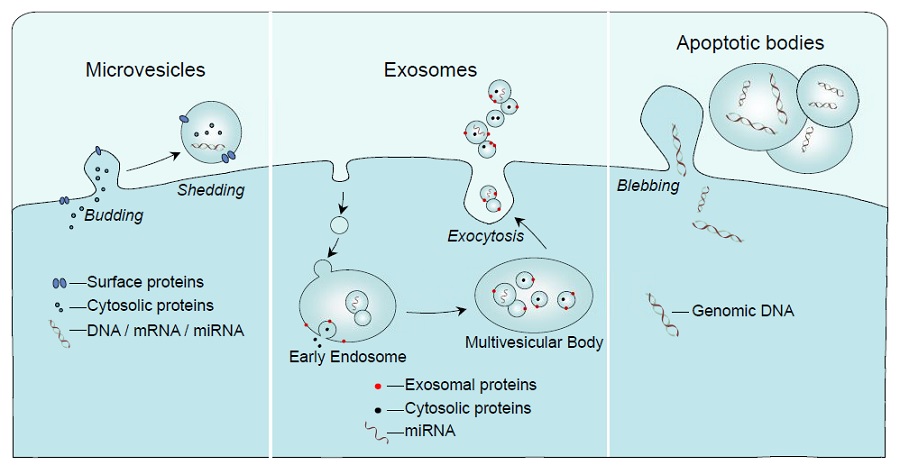
Schematic representation of the mechanisms of formation of microvesicles, exosomes and apoptotic bodies. Reproduced with permission from Journal of Endocrinology 228 R57-R71
Extracellular vesicles (EV) are sub-micron, lipid-encased particles that have been found in all bodily fluids so far tested, including plasma, urine, milk, tears, sweat and semen. They have also been identified in the plant kingdom and in micro-organisms.
The vesicles typically carry a cargo which can include genetic material such as mRNA, genomic DNA and microRNA (miRNA). They may also carry proteins such as growth factors, enzymes and cytokines. Thus, they can be considered as intercellular transport vehicles, with the capacity to influence the behaviour of distant cell types within the body, as well as being important for the transfer of information between individual organisms of the same or separate species.
Until relatively recently, all classes of EV were considered cell debris and not worthy of further study. However, there is now a growing body of evidence to suggest that they have potential as biomarkers or as mediators of disease in a range of different conditions including cardiometabolic disease and diabetes.1
CLASSES OF EXTRACELLULAR VESICLES
Typically, EV are divided into three main populations according to their diameters: exosomes at 50–100nm, microvesicles (MV; microparticles) at 100nm–1μm, and apoptotic bodies at usually greater than 1μm.
Exosomes are produced via an endosomal pathway which involves formation of multivesicular bodies that are trafficked to the plasma membrane, where they fuse to release their exosomal contents. In contrast, both MV and apoptotic bodies are formed by budding or blebbing of the cell membrane to pinch off new vesicles, in a calcium-dependent process (Figure).
This article will concentrate on the measurement and isolation of exosomes and MV.
SEPARATION METHODS
Size ranges of different EVs may overlap, and the markers which are displayed on the outer leaflet of the surrounding membrane may be common to both exosomes and MV. Thus, most isolation methods do not guarantee a pure population of vesicles. With this in mind, and based on the most common protocols involving sequential ultracentrifugation steps, some researchers now refer to each population by the speed at which it is pelleted, rather than referring to exosomes or microvesicles.
'All classes of EV were considered cell debris and not worthy of further study ... there is now a growing body of evidence to suggest that they have potential as biomarkers or as mediators of disease'
There are guidelines available for the collection of blood in particular,2 and several protocols have been published for the sequential isolation of different populations via ultracentrifugation.
In the first step, bodily fluid or culture medium is centrifuged at low speed (1,500–3,000×g (times gravity)) to remove cellular material and debris. The supernatant from this step can then be further centrifuged at 10,000–17,000×g to pellet MV, with a final centrifugation at 100,000×g to pellet exosomes. In addition to exosomes, this population is likely to contain small MV and possibly some lipoproteins. Density gradient ultracentrifugation may be further employed for a purer exosome population.3
In addition, commercially available kits utilise size exclusion chromatography and magnetic separation based on CD9 or CD63, cell surface markers which are exposed on exosomes but not thought to be expressed on MV. Exosomes are directly precipitated from plasma or culture supernatant using these approaches.
However, there is no clear consensus as to the efficacy of these methods in the published literature. For these reasons, it is imperative to characterise the population of interest as fully as possible.
CHARACTERISING EXTRACELLULAR VESICLES
A selection of methods have been utilised for characterisation of EV. Electron microscopy (EM) is considered the gold standard, as it is can give accurate information about the sizes of all classes of vesicle. However, as it is not a quantitative technique and requires specialised expertise and equipment, EM may be of limited appeal. Several other non-optical methods have also been utilised but are limited by the same constraints.
Thus, many researchers have opted for one of a range of optical techniques, of which flow cytometry (FCM) is the most widely reported for MV detection. FCM enables EV phenotyping using fluorochromeconjugated antibodies to determine the parental cell type, and is also quantitative. Small particle size at the limit of detection may be an issue; however, there are a number of sophisticated protocols to eliminate background noise. Newer instruments have lower detection limits and those instruments such as the ImageStream® (Amnis, Seattle, WA, USA) are able to further differentiate between different types of particles in whole blood and plasma.4
Accurate detection of exosomes using FCM should be carried out with caution, as their size is below the limit of detection of many instruments. However, alternative methods are available including nanoparticle tracking analysis and tunable resistive pulse sensing. These techniques enable quantitation of exosomes and small MV, but they have limited capabilities for phenotyping and may require additional time for purification of vesicles before measurement.5
IN CONCLUSION
With increasing evidence for the importance of EV as mediators of inflammation, their potential as biomarkers and their appeal as delivery vehicles for miRNA or other possible therapies to specific target cells, interest in their measurement and isolation has grown exponentially over the last decade. This has led to the availability of an ever-increasing range of instruments and isolation kits, but more established techniques such as ultracentrifugation and FCM should be accessible to most individuals wishing to start exploring this exciting new field.
Charlotte Lawson
Senior Lecturer, Comparative Biomedical Sciences, Royal Veterinary College, London
REFERENCES
1. Lawson C et al. 2016 Journal of Endocrinology 228 R57–R71.
2. Lacroix R et al. 2012 Journal of Thrombosis & Haemostasis 10 437–446.
3. Théry C et al. 2006 Current Protocols in Cell Biology chapter 3 unit 3.22.
4. Headland SE et al. 2014 Science Reports 4 5237.
5. van der Pol E et al. 2010 Journal of Thrombosis & Haemostasis 8 2596–2607.

