Artificial intelligence (AI), a blanket term for several advanced functions undertaken by computers, has already seeped into many aspects of patient care in endocrine surgery. Our specialty focuses on optimising outcomes for patients with thyroid, parathyroid and adrenal disease. This involves accurate diagnostics, assessment of benefits and risks, surgical care with minimisation of morbidity, and follow-up tailored to outcome and risk. AI is already contributing in multiple ways, some in research and others in approved commercial products.
DIAGNOSTICS
- Thyroid nodules may be detected in ultrasound images by computer vision, with features extracted and classified into ‘explainable’ categories (e.g. TI-RADS, the Thyroid Imaging Reporting and Data System). Commercially available ultrasound platforms with this capability include the S-Detect from Samsung.1 Novel deep-learning models as seen in the ThyroNet-X4 Genesis2 and ThyNet3 may automatically classify nodules as benign or malignant, the former using convolutional neural networks and the latter combining three pre-existing AI systems. Real-time image overlay may then guide fine needle aspiration, while cytology slides and molecular tests, such as Veracyte’s Afirma Gene Expression Classifier (GEC) and Genomic Sequencing Classifier (GSC), can be analysed by machine-learning algorithms.4
- In parathyroid disease, machine-learning classifiers may be trained to identify the pathognomonic pattern of classic primary hyperparathyroidism (pHPT) with hypercalcaemia and inappropriately unsuppressed parathyroid hormone (PTH), and to evaluate bloods and urinalysis to exclude alternative diagnoses. AI has even diagnosed those at risk of pHPT from medical notes alone with 86% accuracy.5
- In adrenal diagnostics, radiomics combined with machine learning has shown promise in distinguishing benign from malignant disease.6
Multimodal data integration may be used to assimilate biochemical, imaging and tissue data to give a diagnosis.
PREDICTING PATIENT OUTCOMES
‘Multimodal data integration may be used to assimilate biochemical, imaging and tissue data to give a diagnosis.’
This is an area in which most endocrine surgeons pride themselves: we hope to provide each patient with an understanding of the individualised benefits of surgery, as set against our own operative outcome data. The area is ripe for AI optimisation and most developed in thyroidectomy:
- A deep-learning neural network model trained on the American College of Surgeons National Surgical Quality Improvement Program (ACS NSQIP) data predicted complications and reoperation with 78–97.2% accuracy.7
- Post-thyroidectomy hypocalcaemia may be quantified using a machine-learning model with sensitivity and specificity up to 92 and 90%.8 ‘Super learner’, a cross validation–ensemble approach, predicts hypocalcaemia significantly better than standard multi-logistic regression.9
- Similar models have also been applied preoperatively to predict recurrent laryngeal nerve injury and haematoma, and postoperatively to predict thyroid cancer recurrence.9,10
INTRAOPERATIVE
Over the last century, the focus has shifted from addressing mortality to eliminating morbidity. Thyroidectomy mortality decreased from 40–50% to 0.5% by 1912 (mostly thanks to the influence of Theodor Kocher), and adrenalectomy mortality fell from 30–45% in the early 1900s to 0.2–0.5% in hospital mortality (British Association of Endocrine and Thyroid Surgeons (BAETS) UK Registry of Endocrine and Thyroid Surgery (UKRETS) and ACS NSQIP data respectively).11,12
Morbidity has also declined. Recurrent nerve injury reduced from ~20% in the early 1900s to 3.7–8.6% (for benign and malignant pathology respectively) by the 2000s, and late hypocalcaemia decreased to ~6% in the BAETS UKRETS by 2021.11
These changes occurred before the widespread uptake of technology in which AI may be influential, and were driven entirely by surgical technique, training and specialisation. The volume–outcome relationship reinforces this:13 surgeons with a larger volume of practice and data have more opportunity to hone their operative decision making and techniques.
AI’s intraoperative role may be twofold: helping high-volume surgeons to achieve zero morbidity, and accelerating the learning curve for trainees and low-volume surgeons. With morbidity in high-volume hands as low as <1% recurrent nerve injury, <2% hypoparathyroidism, ~2% persistent hyperparathyroidism and 1% adrenal morbidity, further advances will be more challenging, yet AI may help.
Intraoperatively, AI is seen in two distinct areas:
- It has a role in optimising the output of platforms used and interpreted by the surgeon, such as nerve monitoring, real-time parathyroid imaging and intraoperative PTH testing.
- It supports minimally invasive and robotic procedures, where preoperative imaging may be overlaid to guide anatomy and vascularity, and smart instruments may include activity or force limits.
Nerve monitoring
‘Evidence for AI-assisted technologies (e.g. nerve monitoring, parathyroid assessment and PTH estimation) is mixed, with outcomes still dependent on the surgeon.’
This technology provides information on laryngeal nerve function by applying a small (1–3mA) signal to that nerve and detecting vocal cord movement via sensors on the endotracheal tube. Amplitude and latency are displayed graphically with accompanying sound. The surgeon may stimulate the vagus, recurrent or superior laryngeal nerve via a handheld probe intermittently (intraoperative neurophysiological monitoring, IONM) or continuously (CIONM), the latter requiring the surgeon to attach an electrode to the vagus, delivering a signal each second to show moment-by-moment changes. AI may be used in both modes, particularly CIONM, to track trends, predict impending injury and alert the surgeon.
Parathyroid detection/assessment
The adjunctive technology of near-infrared parathyroid assessment with a probe (PTeye) or camera (Fluobeam) may confirm parathyroid identification and, via autofluorescent contrast (indocyanine green, ICG), may map vasculature and assess perfusion. Both systems include AI software to optimise their function, although limitations remain currently: subsurface glands are not detected, adenomas vary in signal, and function cannot be reliably predicted.
Point-of-care blood tests
Intraoperative PTH estimation may be used to predict cure of hyperparathyroidism by the surgeon. PTH dynamics are generally predictable and interpretation is therefore straightforward in most cases. However, subtleties may mislead the unwary surgeon. AI has been shown to improve accuracy and decision support.
Evidence for AI-assisted technologies (e.g. nerve monitoring, parathyroid assessment and PTH estimation) is mixed, with outcomes still dependent on the surgeon.
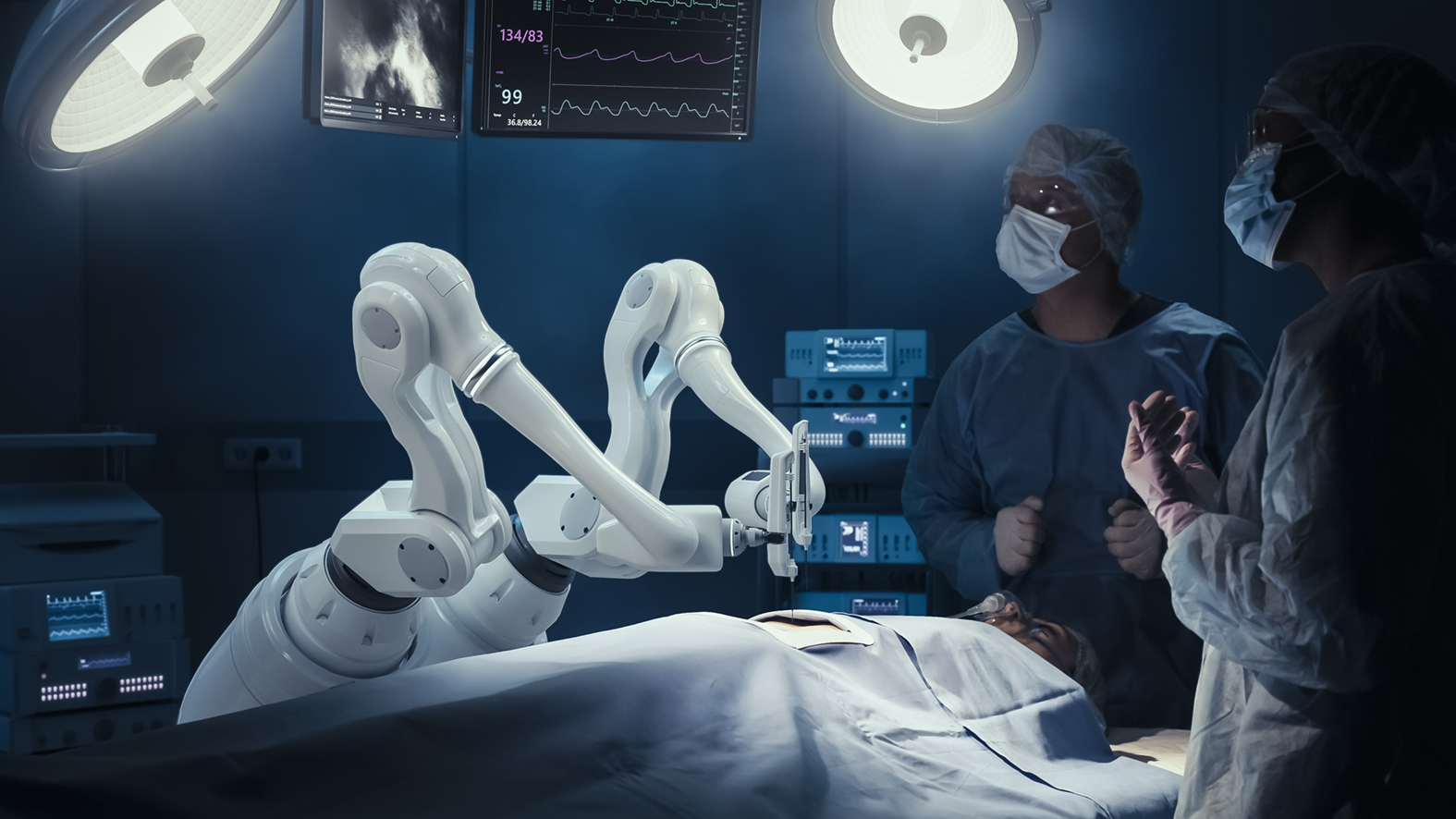
Robotic surgery
The application of robotic surgery is growing but has limitations. Laparoscopic or retroperitoneoscopic adrenalectomy remains the gold standard, and, while switching to robotic adrenalectomy is therefore readily achieved, the benefits are unproven. In neck endocrine surgery, access is problematic: transaxillary, sub-mammary and transoral approaches all have limitations, in part due to the size of the robotic instruments. Lower profile instruments and smart instruments (containing haptic feedback and enhanced integrated tools, such as nerve monitoring) have promise for the future.
IN CONCLUSION
AI is already present in key areas of endocrine surgery, particularly in diagnostics and outcome prediction. Currently, outcomes remain surgeon-dependent, with mixed data on intraoperative adjuncts. However, as AI improves and refines, its role in surgical care is also likely to increase.
AIMEE DI MARCO
Endocrine Surgeon, Hammersmith Hospital and Imperial College and NHS Trust, London
REFERENCES
1. Barczyński M et al. 2020 Gland Surgery https://doi.org/10.21037/gs.2019.12.23.
2. Wang X et al. 2025 Scientific Reports https://doi.org/10.1038/s41598-025-86819-w.
3. Peng S et al. 2021 Lancet Digital Health https://doi.org/10.1016/S2589-7500(21)00041-8.
4. Wong CM et al. 2023 Journal of Pathology Informatics https://doi.org/10.1016/j.jpi.2023.100309.
5. Greer ML et al. 2022 Head & Neck https://doi.org/10.1002/hed.26970.
6. Ferro M et al. 2025 Therapeutic Advances in Urology https://doi.org/10.1177/17562872251352553.
7. Tsutsumi K et al. 2023 Clinical Otolaryngology https://doi.org/10.1111/coa.14066.
8. Muller O et al. 2024 Annals of Surgery https://doi.org/10.1097/SLA.0000000000006480.
9. Seib CD et al. 2021 American Journal of Surgery https://doi:10.1016/j.amjsurg.2020.11.055.
10. Ahmad M et al. 2024 Arχiv https://doi.org/10.48550/arXiv.2410.10907.
11. BAETS 2021 Sixth National Audit Report https://baets.org.uk/wp-content/uploads/2024/08/BAETS-Sixth-Audit-Report-FINAL.pdf.
12. Gupta PK et al. 2011 Surgical Endoscopy https://doi.org/10.1007/s00464-010-1256-y.
13. Stavrakis AI et al. 2007 Surgery https://doi.org/10.1016/j.surg.2007.09.003.

